

Looking Back: Me and My Bubble
“The bubble is trying to keep me from writing about the bubble”
I am, right now, experiencing a phenomenon that most of you mercifully will never have to endure. I have a gas bubble in my right eye that has been there since April 26th and it is shrinking, ever so slowly, in size. It is driving me nuts but I am eternally grateful that it is there. I must add that it is pretty damn awkward and frustrating trying to type while being obstructed this way.
Ironically, I spent many years of my life maneuvering a bubble in order to level my survey instrument and now, I have one in my eye that is acting exactly the same. It appears to stays level despite its spherical shape, as I tilt my head one way or the other. As of June 15th it is still obstructing the bottom half of my eye. Fun fact: I can actually make my bubble bobble.
A recent follow up visit to Dr. Jeff Mann at Rocky Mountain Optometry about my bubble eye repair progress revealed that the bubble is in fact not in the bottom of my eye, but at the top. Simple eye dynamics that I had forgotten. An image passing through the eye is inverted and the brain then reverses it. All this bubble monkey business came about from what it termed a macular hole repair and an eye degeneration journey that I have been on for over 4 years.
Perhaps a little background on all this gas bubble business is in order, so what say we back up this retinal bus to January 1st, 2022. On New Year’s Day, of all days, I turned on the TV and could not seem to be able to focus on the screen. There was an ominous gray spot obstructing part of my left eye. It didn’t look like it was going to go away any time soon so my ADHD brain kicked in big-time into panic mode, as I contemplated, “was this a retinal detachment”? Incidentally, these detachments or tear incidents require immediate attention, as you can lose your sight if they are not repaired within a couple of days. So I was pretty much fully wired about it all.
I thought perhaps I should head to the hospital, but then realized that there was nothing that they could do diagnostically. I then had an idea hatched out of desperation and sent a message to Rhonda Bond, someone I trusted and who was then an employee with Rocky Mountain Optometry but has since retired. I shared with her my concern in the hopes that she would spot the message even though we are not friends on Facebook. It was not 45 minutes later that she messenger messaged me back that she had called Dr. Jeff Mann, co-owner of the business and a top notch optometrist that has been working in a field he absolutely loves for 34 years. She informed me that he would be calling me in the next little while.
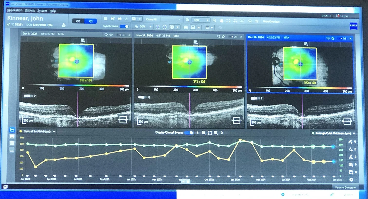
Jeff did, in fact, call from his place in Cowley, which was, like here at the time, experiencing minus 30 Celsius temperatures and a nasty east wind blowing. Jeff said he would try to get in, but wasn’t sure he’d make it and if he did I was to meet him at the office downtown Blairmore at two o’clock. True to his word he made it there and in advance had contacted one of the girls that work at the office to open it up and fire up the computers. On New Year’s Day!! My eye was photographed using OCT (Optical Coherence Tomography) and Mann assessed this imagery and revealed that it was not a retinal detachment but in fact, the beginnings of wet macular degeneration.
A simple definition of this term is probably in order here: “Wet macular degeneration is a serious, fast-progressing eye condition that happens when abnormal, fragile new blood vessels grow under the retina and leak fluid and blood, causing the macula—the part of the eye responsible for sharp, straight-ahead vision—to swell and scar, which leads to sudden vision loss.”
So unlike dry macular, which is progressive over sometimes a long period of time and cannot be treated directly, wet macular has a treatment option.
Within two weeks of my diagnosis I was travelling to the Dr Michael Johnson private eye care clinic in Lethbridge; for the first of a series of what will be pretty much a lifetime of injections in my left eye. The injections are designed to keep the degeneration at bay (not a cure) and generally happen every 6 to 7 weeks. While this might sound onerous, I am eternally grateful that this process was developed because there are thousands of us out there that are developing wet macular.
The technique was discovered accidentally by physicians using a particular chemistry to restrict blood vessels growing with stomach cancer. Cancer patients began reporting some improvement in their eyesight. The light came on and now that chemistry, called anti-VEGF (anti-vascular endothelial growth factor) is used to control wet macular and is administered by specially trained ophthalmologists across Alberta. The injection process is very simple and conducted with every care, using freezing, and a lot of flushing to keep your eye healthy and safe.
Fast forward then to January 2026 when a standard scan that day prior to a scheduled injection revealed that the right eye appeared to have moved in the same direction as the left. It was one that they had been watching closely since 2022 as it was very close to going wet. Injections were started in both eyes that day but on the very next scheduled visit the scan revealed something a little more concerning. It is never good when you hear an ophthalmologist say,”oh that’s not good.”
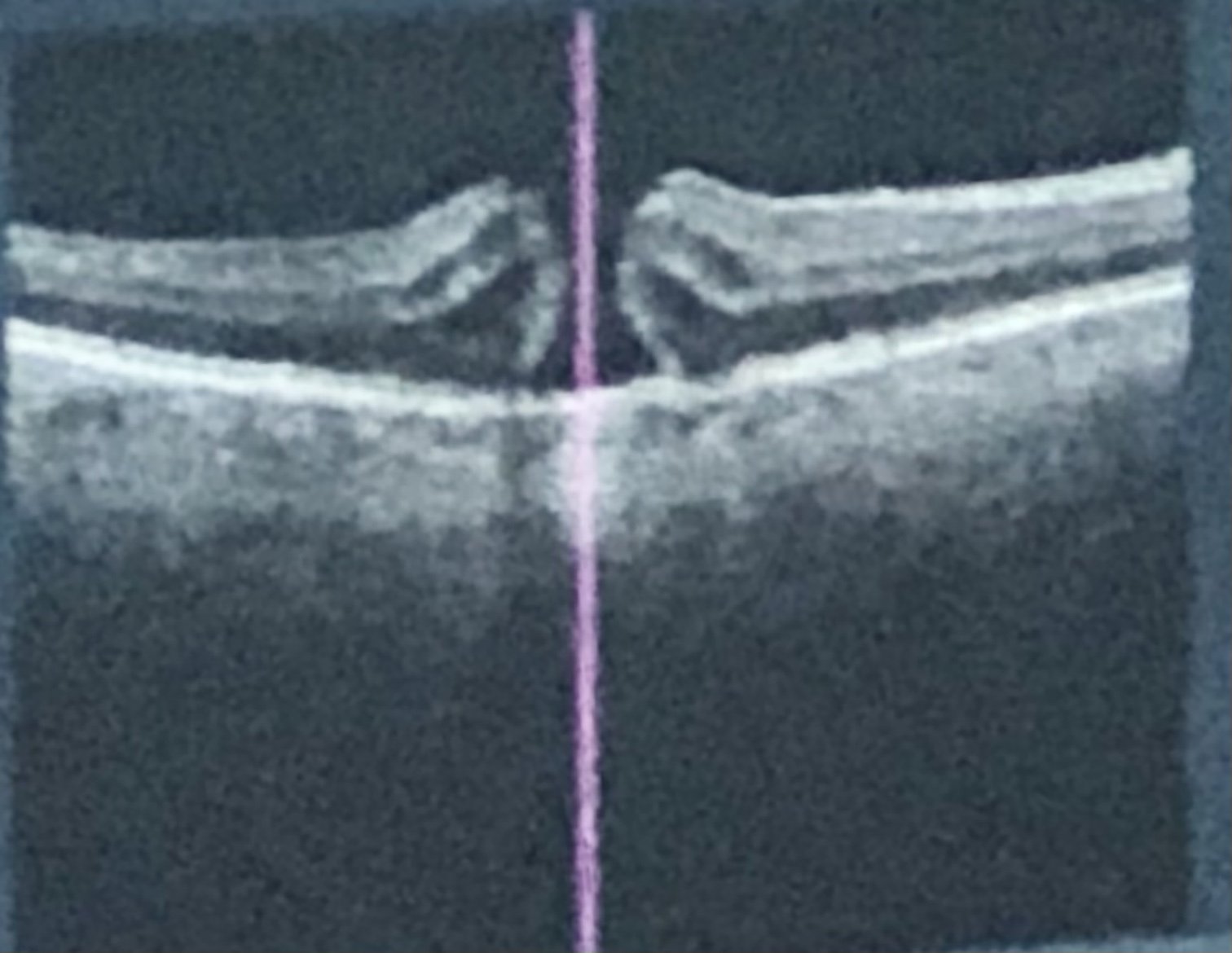
What he saw in fact was a macular hole, a small, round opening in the macula—the tiny, light-sensitive area at the very center of your retina. It is responsible for sharp, detailed central vision so a hole in the area causes blurriness and wavy vision. I had been experiencing a lot of that at the time which made driving a bit dicey. And no matter the reading glass’ strength or magnifying glass maneuvers, I could not, for the life of me, focus properly. It was beyond frustrating. The decision was made then not to inject the right eye because this issue required surgery by a specialist in Calgary, so best not to disturb it prior.
Johnson booked me, in late March, into the Mitchell Eye Centre in Calgary where I was put through a battery of tests in no less than four different rooms, with many sophisticated devices, before I eventually saw a specialist. It was agreed then that we do the surgery and in the process remove the developing cataract as well. Two weeks later I had to present myself for an advance surgical assessment and yet more rooms and optical wizardry. The centre is like an assembly line but very professional and thorough.
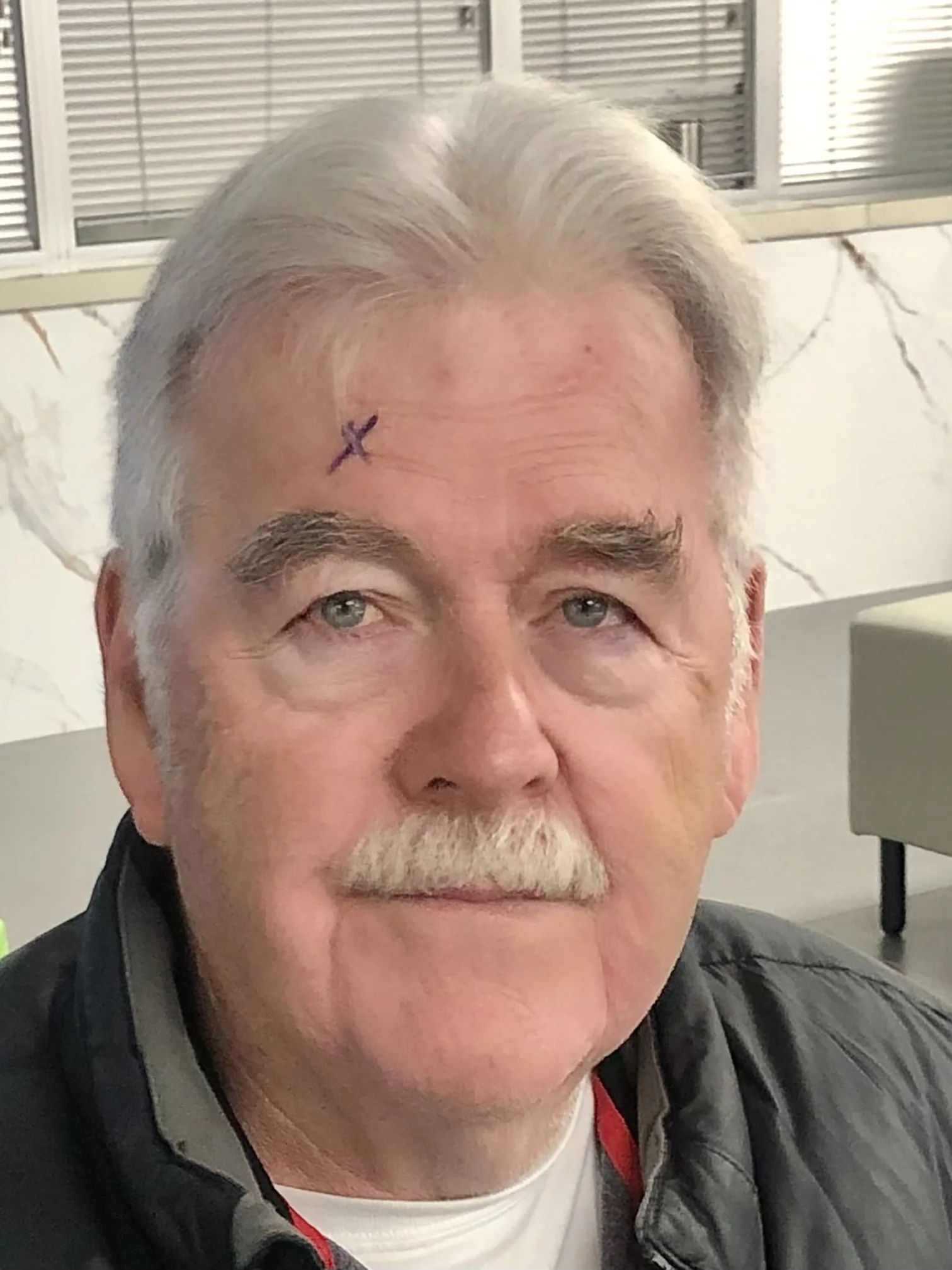
On April 26, at the Old Holy Cross Hospital optical wing, I met the specialist Dr. Jessica Ruzicki, for the scheduled surgery. Like the Mitchell Centre, the Holy Cross moves you through at a steady pace where numerous patients have different issues dealt with. I sat and waited with a black X over my right eye and finally entered a room that had no less than 15 reclining optician’s chairs, many of them containing patients awaiting pre-treatment protocols. I was checked over by the anesthesiologist, my blood pressure monitored, and given yet another round of drops. I was then given an injection underneath my eye that froze the right side of my face. Eventually I was moved to surgery where I was treated with a nice warm blanket. I lay there with arms crossed and was conscious through the whole procedure. There were a lot of funny sounding devices that were operating out of my periphery which intrigued my ADHD curious mind but I am glad I could not see them.
Dr. Ruzicki proceeded to remove the cataract and then repair the macular hole, after which a neutral gas was injected into my eye. Its purpose is to hold the macular repair in place. Then it was back to my hotel room, as I had to report to the Mitchell Centre the next morning, bright and early, for a quick inspection apres surgery. I returned to my room at the Carriage House where I was met later by a representative of a company called Calgary Vitrectomy Recovery Equipment, who came to my room armed with several devices that I had been advised to rent. Vitrectomy is defined as a surgical procedure to remove all or part of the “vitreous humour” – (clear gel substance filling the centre of your eye. It was done in my case in order to access and repair the retina at the back of the eye. Well yikes you say. That all sounds pretty dicey, but not to worry. It is all standard procedure and done every day with success.
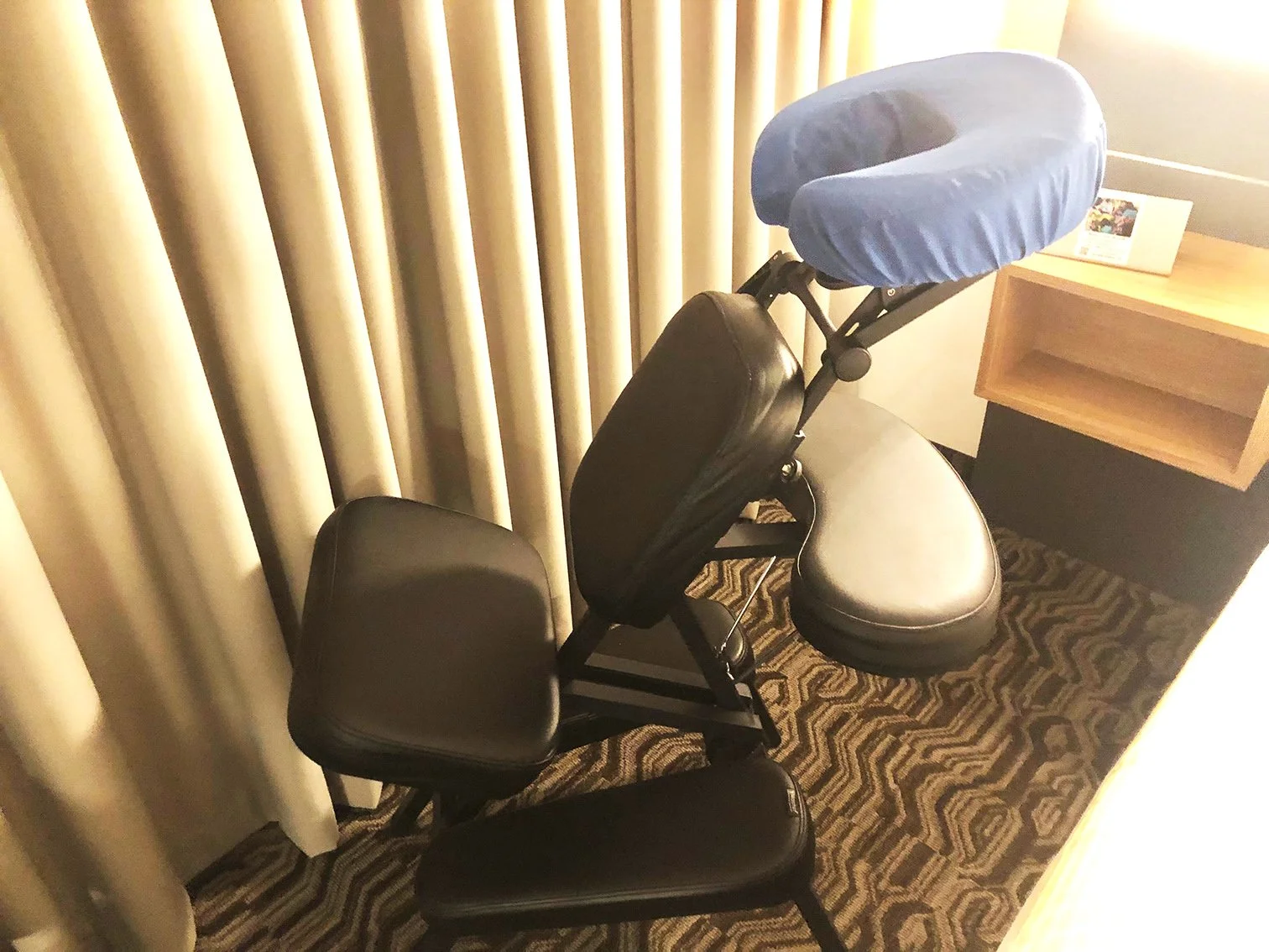
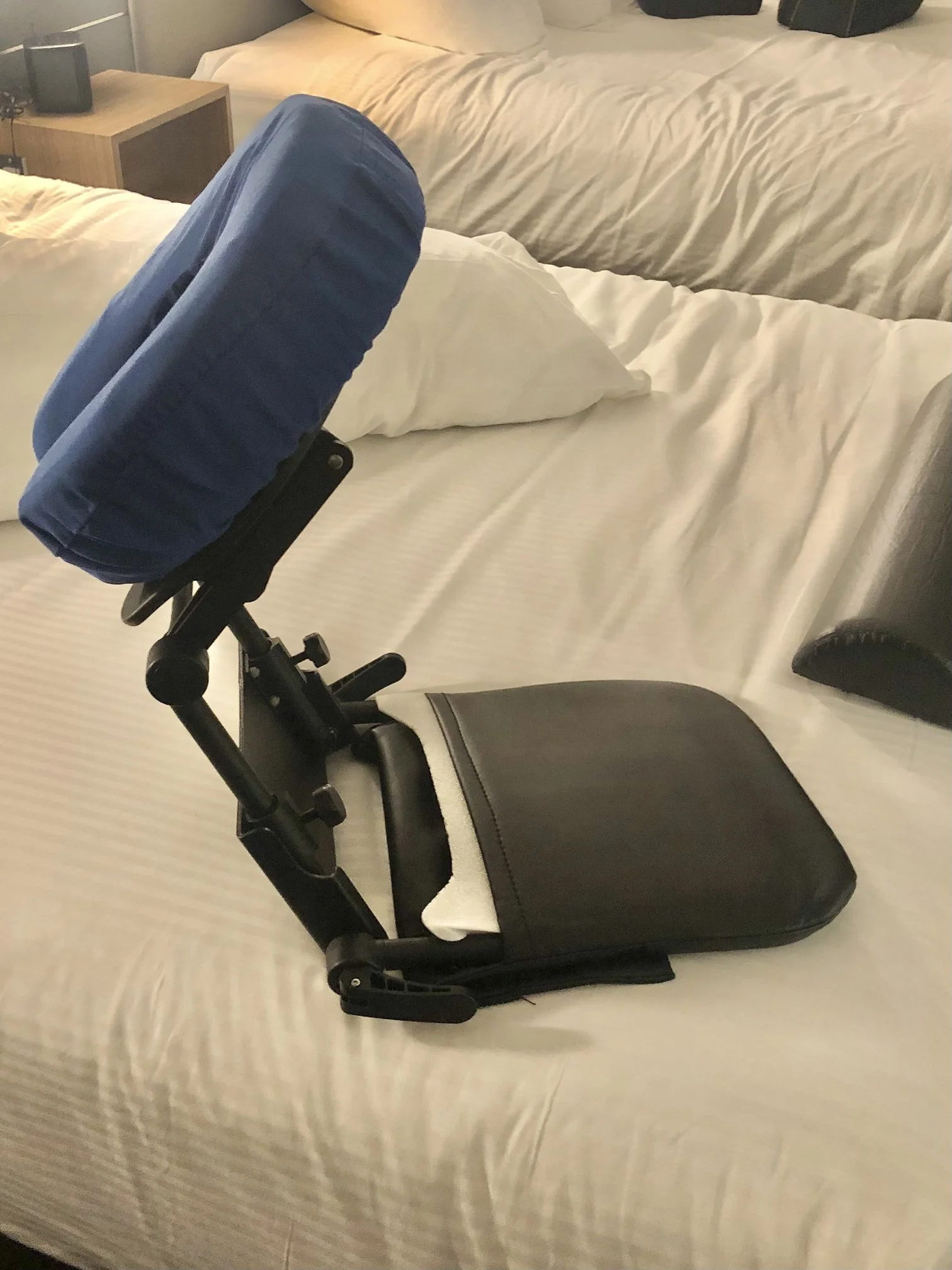
The “Retinal Care Specialist, named Cam, spent over an hour setting up and adjusting pieces of a rented package that were designed to help me through the first part of an awkward recovery. It included a special chair with a rest area for my arms, pads for my knees and what I call a face doughnut up front. It is a U-shaped cushion that one can put one’s face into. I could see through it and keep, as required; my bubble loaded eyeball, face down. This face down protocol was an absolute must for the first week in order that the bubble, which uses its volumetric pressure, hold in place the repair at the back of the eye.
There was a second smaller doughnut device provided that was mounted to a stand that could sit at a table so you could have some variety in your day. The device could be flattened out so that you could lay face down in bed. The first night with the devices, in the hotel room, was sort of a disaster. There was a lot of shifting around and trying whatever position I could to stay face down. I can tell you laying on your face in a bed looking into a face planter is more than awkward. It’s downright uncomfortable.
Author’s Note: My bubble story is a long one so stay tuned next week for Part Deux of “Me and My Bubble”. Or should it be, “My Bubble and Eye”.